The 2 valves that separate the ventricles from the atriaThe atria are two of the four cavities of the heart. There is a right atrium and a left atrium. They are like reservoirs for the right and left ventricules. are called atrioventricular valves. The tricuspid valve is located on the right side and the mitral valve is on the left.
A different architecture
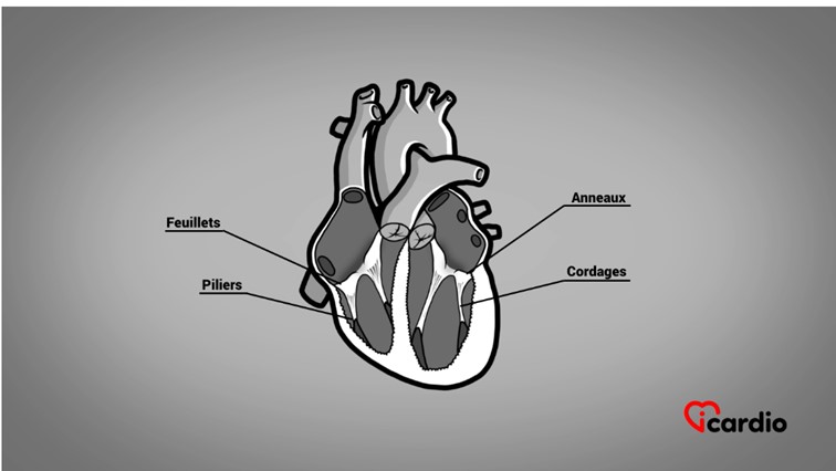
Each of these valves is made up of a fibrous ring from the cardiac skeleton, with leaflets, cords, and muscular pillars placed inside the lower part of their respective ventricle.
These elements constitute an actual valve system, in comparison to the other 2 valves of the heart, the aortic and pulmonary valves.
The mitral valve apparatus
The mitral valve can be compared to a sailboat. We have the mast, the ropes that connect the sail to the structures, and the sail itself.
The 2 leaflets are attached all around the fibrous ring. The free edges of the leaflets are not smooth; they form extensions similar to a short net. From those, ropes are formed and tied to the muscular pillars at the bottom of the left ventricle.
The Valves Are Sealed
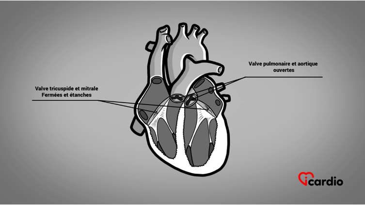
Their configuration is such that the valves are hermetically sealed.
When the ventricles contract, the pressure generated by the muscle propels the heart’s contents upwards. This impulse closes the mitral and tricuspid valve leaflets. The cords prevent them from folding back into the atrium, shutting them tightly. Bloodblood Blood comprises red cells, white cells, platelets, and plasma. Red cells carry oxygen and carbon dioxide. White cells are our defense system. Platelets prevent bleeding. Plasma is the liquid component of blood. It contains water, salts, has no choice but to be expelled through the pulmonary valve on the right and the aortic valve on the left.
Resistance to High Tension on the Leaflets
A strong thrust strikes this barrier with each contraction. This is more specifically the case for the mitral valve, due to the higher pressures on the left than on the right.
One can imagine the situation in patients with systolic pressures over 200 mmHg. The same tension is exerted on the mitral valve, and particularly on its cords, which are firmly anchored to the muscular pillars. This is the centerpiece of a highly resistant architecture.